X-Ray Computerized Tomography (Micro CT 1273)
Price
| Price | |
| Instrument usage per hour | 2450 |
| Instrument usage per hour half hour | 1225 |
| Quantitative analysis per sample | 1750 |
| Use heating and cooling stages / temp / sample | 1750 |
| Use compression or tensile testing stages / sample | 1750 |
| Instrument usage per hour (for authorized user only) | 1960 |
| Program analysis per hour (for authorized user only) | 875 |
| Training fee per hour | 1000 |
| Price | |
| Instrument usage per hour | 2800 |
| Instrument usage per hour half hour | 1400 |
| Quantitative analysis per sample | 2000 |
| Use heating and cooling stages / temp / sample | 2000 |
| Use compression or tensile testing stages / sample | 2000 |
| Instrument usage per hour (for authorized user only) | 2240 |
| Program analysis per hour (for authorized user only) | 1000 |
| Training fee per hour | 1000 |
| Price | |
| Instrument usage per hour | 3150 |
| Instrument usage per hour half hour | 1575 |
| Quantitative analysis per sample | 2250 |
| Use heating and cooling stages / temp / sample | 2250 |
| Use compression or tensile testing stages / sample | 2250 |
| Instrument usage per hour (for authorized user only) | 2520 |
| Program analysis per hour (for authorized user only) | 1125 |
| Training fee per hour | 1000 |
| Price | |
| Instrument usage per hour | 3500 |
| Instrument usage per hour half hour | 1750 |
| Quantitative analysis per sample | 2500 |
| Use heating and cooling stages / temp / sample | 2500 |
| Use compression or tensile testing stages / sample | 2500 |
| Instrument usage per hour (for authorized user only) | 2800 |
| Program analysis per hour (for authorized user only) | 1250 |
| Training fee per hour | 1000 |
X-Ray Computerized Tomography (Micro CT 1173)
Bruker
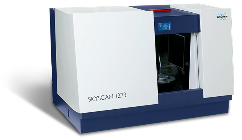
Model : SKYSCAN 1273
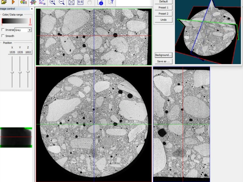
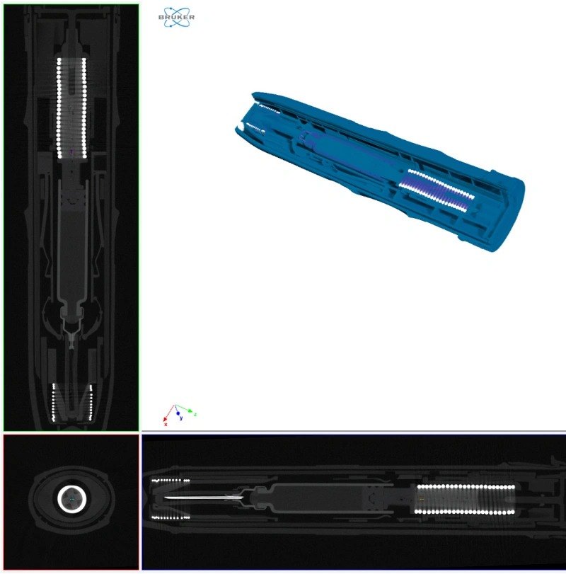
เครื่องถ่ายภาพรังสีเอ็กซ์เรย์ ด้วยคอมพิวเตอร์ (X-Ray Computerized Tomography) หรือ Micro CT หรือ X-Ray Microscopy (XRM) เป็นเครื่องมือวิทยาศาสตร์ที่ใช้เทคนิคการถ่ายภาพด้วยรังสี X-ray ที่มีความละเอียดสูง ซึ่งใช้คอมพิวเตอร์ในการประมวลผลสร้างภาพตัดขวาง 2D และ 3D จึงสามารถดูภาพโครงสร้างและรายละเอียดภายในของตัวอย่างได้ นอกจากดูภาพสามมิติได้แล้ว ภาพจากเครื่อง Micro CT สามารถคำนวณค่าพารามิเตอร์ต่าง ๆ ของตัวอย่างได้ เช่น Total VOI volume, Object volume, Porosity, BMD และอื่น ๆ
Feature
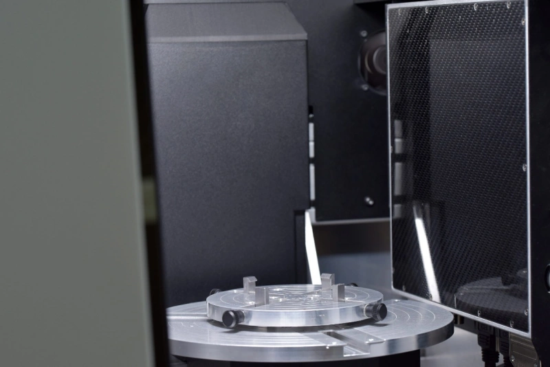
เครื่อง Micro CT BRUKER SKYSCAN 1273
- X-ray source: 40-130 kV, 39 W
- Active pixel CMOS flat-panel, 6 MP (3072 x 1944)
- Resolution : 3 micron (Minimum Resolution)
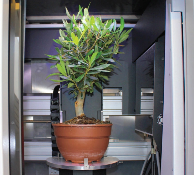
- Object size: 250 mm diameter, 250 mm height
- Stages for Tensile & Compression Testing
Application
- สร้างภาพ 3D จากการแสกนด้วย X-ray เพื่อดูโครงสร้างภายในของตัวอย่าง เช่น รูพรุน, รอยแตก, ลักษณะของเนื้อวัสดุ, ลักษณะของกระดูก หรือเนื้อเยื่อของสิ่งมีชีวิต (ที่ไม่มีชีวิตแล้ว)
- คำนวณค่าพารามิเตอร์ต่างๆ ได้ เช่น Total VOI Volume, Object volume, Closed Porosity, Open Porosity, Total Porosity, Bone Parameter เช่น BMD, BV/TV, Tb.Th
Sample
- Material เช่น หิน, ซีเมนต์, แบตเตอร์รี่, พอลิเมอร์, ชิ้นส่วนอุปกรณ์อิเล็กทรอนิกส์, วัสดุทางทันตกรรม, โลหะ
- Life Science เช่น กระดูก, ฟัน, สิ่งมีชีวิตทางชีววิทยา (แบบไม่มีชีวิตแล้ว)
ลักษณะรูปร่างของตัวอย่าง : ตามธรรมชาติของตัวอย่าง, ทรงกระบอก, ลูกบากศ์
ขนาดของตัวอย่าง : ยิ่งมีขนาดเล็ก จะเข้าใกล้ X-ray ได้มากขึ้น ทำให้ได้ความละเอียดภาพที่ดี จึงแนะนำให้ขนาดเล็กที่สุดเท่าที่ขอบเขตเงื่อนไขที่ต้องการศึกษา
Staff

นางสาวจิราพัชร ประเสริฐทรัพย์
เจ้าหน้าที่บริการวิทยาศาสตร์ P7
Contact
นางสาวจิราพัชร ประเสริฐทรัพย์
- E-mail : Jirapat.pr@chula.ac.th
- Tel. : 02-218-8107
