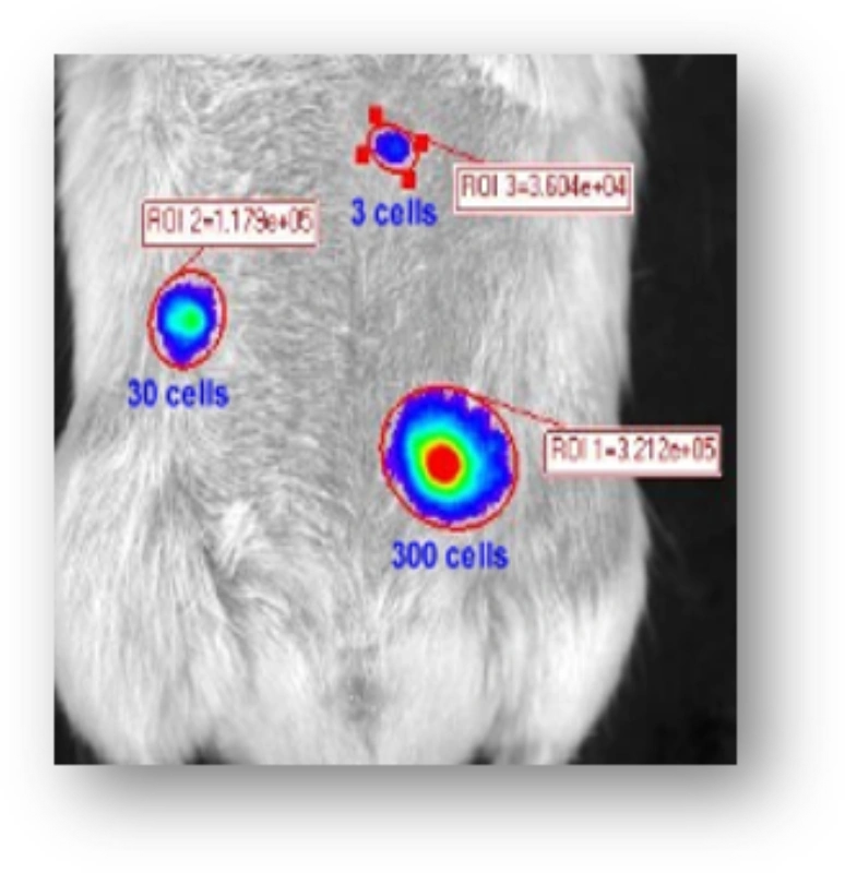
In Vivo Imaging – IVI (IVIS Spectrum)
Price
| Price | |
| Imaging per hour | 1300 |
| Imaging per hour 1st hour (Authorized user) | 700 |
| Imaging per hour next hour (Authorized user) | 100 |
| Price | |
| Imaging per hour (Other University) | 1820 |
| Imaging per hour 1st hour (Authorized user) (Other University) | 980 |
| Imaging per hour next hour (Authorized user) (Other University) | 140 |
| Price | |
| Imaging per hour (Government) | 2080 |
| Imaging per hour 1st hour (Authorized user) (Government) | 1120 |
| Imaging per hour next hour (Authorized user) (Government) | 160 |
| Price | |
| Imaging per hour (Enterprise) | 2340 |
| Imaging per hour 1st hour (Authorized user) (Enterprise) | 1260 |
| Imaging per hour next hour (Authorized user) (Enterprise) | 180 |
In Vivo Imaging
PerkinElmer
Model : IVIS Spectrum
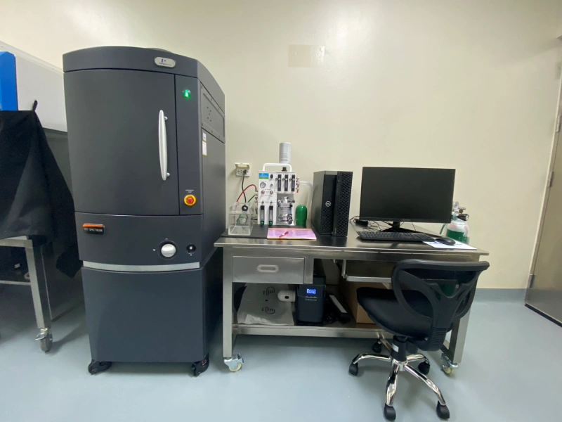
The IVIS® SpectrumCT expands upon the versatility and advanced optical feature sets of the IVIS and Maestro™ platforms integrated with low dose microCT to support longitudinal imaging. The IVIS SpectrumCT enables simultaneous molecular and anatomical longitudinal studies, providing researchers with essential insights into complex biological systems in small animal models. The constant horizontal gantry motion and the flat panel detector provide unparalleled performance for low-dose imaging and automated optical and microCT integration. The stable revolving animal platform table rotates 360° to acquire full 3D data. Multiple animals can be scanned simultaneously while maintaining an average dose per scan at about 13mGy, with a scanning and reconstruction time of less than a minute. Optical and microCT modalities can also operate independently.
Feature
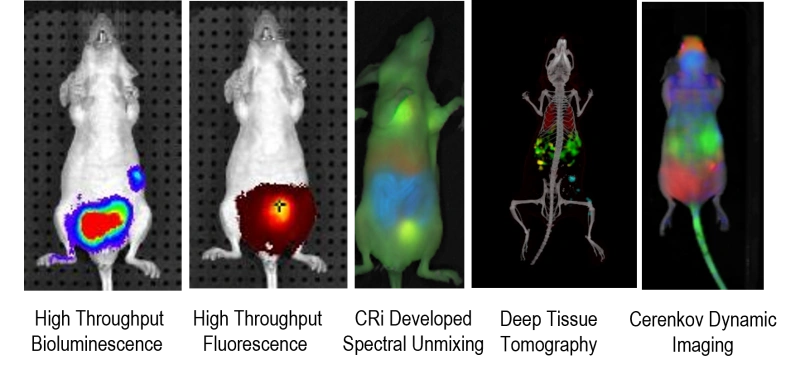
- สามารถตรวจวัดสัญญาณภาพได้ทั้งแบบ 2D/3D optical Fluorescence และ Bioluminescence
- ตัวเครื่องประกอบด้วย กล้อง CCD ชนิดความไวสูง
- ให้ความละเอียดของภาพ 2048 x 2048 pixels มีขนาดของ pixels 5 μm
- มีค่า Quantum Efficiency >85% ที่ช่วง 500-700 nm และ >30% ที่ 400-900 nm
- สามารถถ่ายภาพ Minimum Field of view 3.9×3.9cm และขนาด Maximum Field of view 23×23 cm
- มีค่า Minimum image pixel resolution 20 μm
- มีค่าสัญญาณรบกวน(Read Noise)<3 electrons for bin = 1,2,4; และ <5 electrons for bin=8,16
- มีค่าสัญญาณในที่มืด (Dark Current) < 100 electrons/s/cm2
- มีค่า Minimum Detectable Luminance < 70 photons/s/sr/cm2
- มีชุดเลนส์ขนาด 50 mm สามารถปรับความกว้างของรูรับแสงได้ ตั้งแต่ f/1-f/8
- สามารถปรับกำลังขยายได้ 1.5x, 2.5x, 5x และ 8.7x
- มี Filter สำหรับ excitation filter จำนวน 10 อัน (30 nm bandwidth)
- มี Filter สำหรับ emission filter จำนวน 18 อัน (20 nm bandwidth)
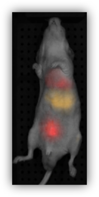
- มีระบบ Spectrum unmixing ช่วยลดการเกิด auto fluorescence และรองรับการถ่ายภาพแบบ multispectral fluorescence
- ใช้ระบบ Laser Galvanometer ในการสร้างภาพพื้นผิวของตัวอย่างแบบ 3 มิติ
- มีระบบ excitation ด้านบนแบบ reflectance illumination และด้านล่างของตัวอย่าง พร้อมแท่นที่สามารถเคลื่อนที่ได้ในแกน X-Y เพื่อเพิ่มความไว และความแม่นยำในการวิเคราะห์เชิงปริมาณสำหรับตัวอย่างที่อยู่ลึกลงไปในสัตว์ทดลองได้อย่างแม่นยำยิ่งขึ้น
Application
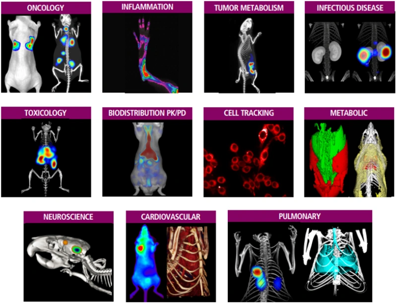
ใช้ในการทำภาพและวิเคราะห์สัญญาณเรืองแสงจากสัตว์ทดลองขณะยังมีชีวิตที่ให้สัญญา Fluorescence หรือ Bioluminescence ซึ่งสามารถประยุกต์ใช้งานในงานวิจัยทางวิทยาศาสตร์การแพทย์ได้อย่างหลากหลาย เช่น การศึกษาเซลล์เนื้องอก การศึกษาด้านภูมิคุ้มกันของเซลล์ ยีนบำบัด การศึกษาเกี่ยวกับการส่งถ่ายของตัวยา(Drug delivery) ทำให้สามารถเก็บข้อมูลจำนวนมากได้อย่างต่อเนื่องในสัตว์ทดลอง ลดจำนวนสัตว์ทดลองที่ต้องใช้ต่อการทดลอง ใช้ศึกษาผลของยา ชีววัตถุ เซลล์ ต่อการเจริญเติบโตของมะเร็ง patient-derived cancer organoid สามารถใช้วัดปริมาตร และติดตามการแพร่กระจายของมะเร็ง ใช้ในการพัฒนาการรักษาแบบ immunotherapy-vaccine, antibodies ติดตามผล gene therapy products และการศึกษาระบบขนส่งตัวยา เป็นต้น
Sample
สัตว์ทดลอง ประเภทหนู Mice หรือ Rat
Staff

สพ.ญ.จีรประภา ดวงบุผา
สัตวแพทย์ P7
Contact
Email : Jeeraprapha.d@chula.ac.th
Tel. : 0-2218-9991
